Indications for ORIF include all fracture patterns that result in hip joint instability of incongruity, bone/soft tissue incarcerated within the joint, or in situations in which non-operative treatment is not a satisfactory option, total hip arthroplasty of percutaneous fixation should be a considered approach.Although the incidence, types, and radiological outcomes of simultaneous ipsilateral pelvic ring and acetabular fractures have been reported, there have been no reports on factors that may affect the quality of acetabular fracture reduction. Prolonged traction (4 to 12 weeks) if fracture indicates surgery, but the patient is not a surgical candidate.
Acetabular fracture full#
Progress to full weight bearing by 6 to 12 weeks (once adequate fracture healing). Begin with foot-flat partial weight bearing (<10kg) radiographs obtained weekly for the first 4 weeks. Once pain allows, immobilization should follow. įractures treated non-operatively require bed rest initially for comfort. If the CT scan shows no fracture lines involving this area, the fracture can be a candidate for non-operative management displacement cannot exceed 2 mm. Evaluation involves scanning from the vertex to 10mm inferiorly. This system divides fractures into five elementary types and five associated fracture patterns.īoth column with secondary congruence (no traction)Įvaluation of the vertex (the most superior portion of the roof) on CT scan can help identify fractures that are amenable to non-operative management. īy far the most used and most useful classification for acetabulum fractures is that of Letournel. Any evidence of hip subluxation means hip instability.
Acetabular fracture manual#
The patient is supine with the hip extended and in neutral rotation, then the hip is flexed to 90 degrees, and a manual force is applied while taking plain films. Dynamic stress X-rays can also be obtained to evaluate for hip stability, usually when there is a fracture-dislocation involving the posterior wall. 2D images are better used for evaluation of the fracture patterns, while 3D imaging may help less experienced surgeons.

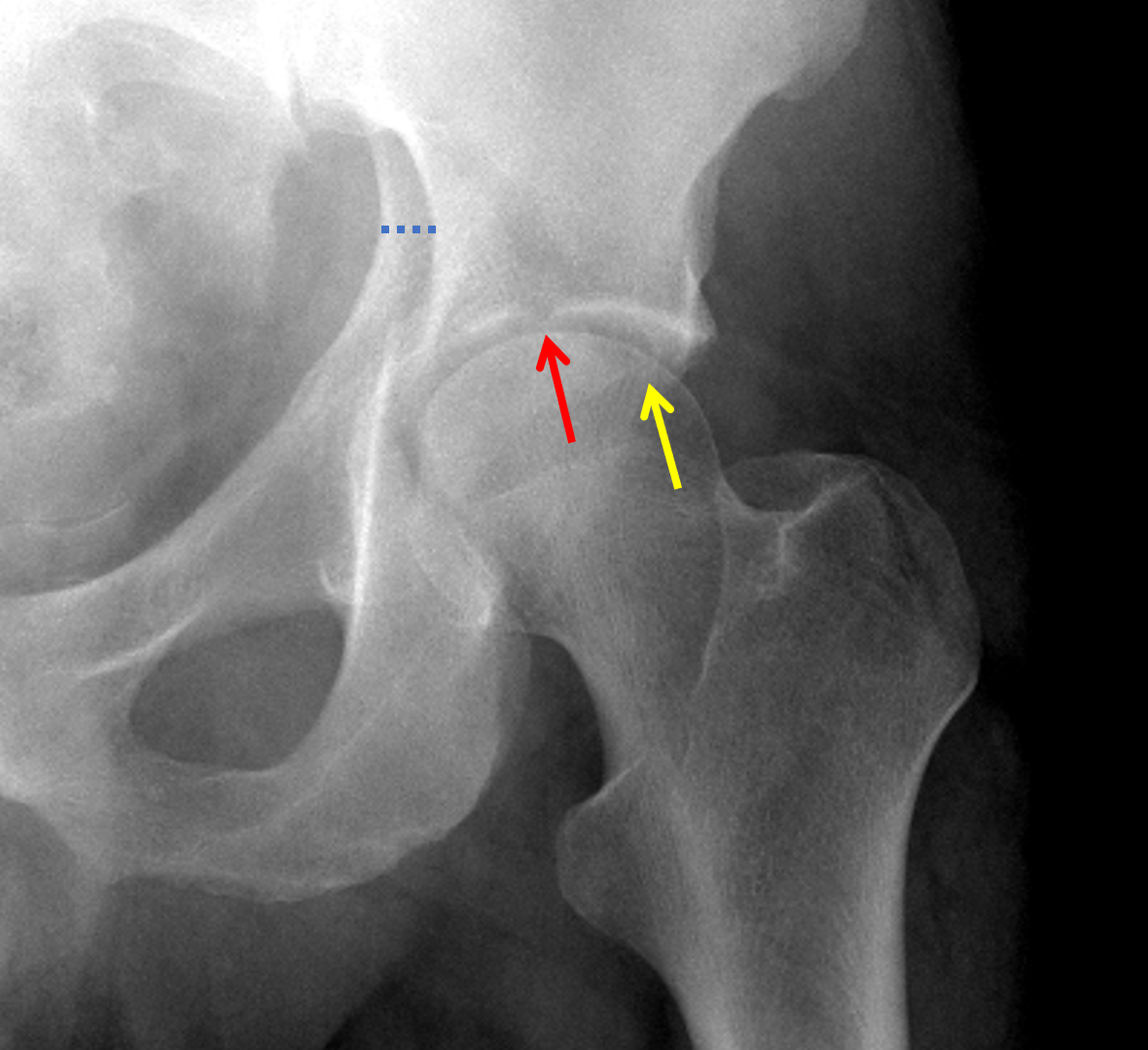
CT axial images are superior to plain radiographs in evaluating the following in acetabulum fractures: Extent and location of acetabular wall fractures, presence of intra-articular fragments, the orientation of fracture lines, the identification of additional fracture lines, rotation of fracture fragments, the status of posterior pelvic ring, and marginal impaction. The advent of CT scans has made the diagnosis and classification of acetabulum fractures much easier. The lateral limb represents the inferior aspect of the anterior wall and the medial limb forms from the obturator canal and the anterior inferior portion of the quadrilateral surface. The teardrop represents a radiographic finding and is not a true anatomical structure. The ilioischial line represents the posterior column, which is a tangency of the quadrilateral surface. The iliopectineal line represents the anterior column and is made up of the pelvic brim and sciatic buttress and greater sciatic notch. The posterior column begins at the superior aspect of the greater sciatic notch, contiguous with greater and lesser sciatic notches, and includes the ischial tuberosity. The anterior column contains the anterior half of the iliac wing that is contiguous with a pelvic brim to superior pubic ramus and anterior half of the acetabular articular surface. The acetabulum divides into an anterior and posterior column. One can visualize the articular surface as an inverted Y with a thick strut of bone connecting it to the sacroiliac (SI) joint, known as the sciatic buttress. Blood supply to the internal surface comes from the fourth lumbar, iliolumbar, and obturator arteries. Blood supply to the external surface is via the superior gluteal artery, inferior gluteal artery, obturator artery, and medial femoral circumflex. The superior portion of the acetabulum articular surface has the name of the weight-bearing dome. The innominate bone forms from the pubis, ischium, and ilium at the triradiate cartilage. Among the most significant advancements has been the advent of percutaneous fixation of certain fracture types. Little has changed since Letournel and Judet’s landmark paper in 1993, and many of their findings remain the “gold standard” for treatment today.

There has been an increase in the number of acetabulum fractures resulting from a fall of fewer than 10 feet, likely due to the rise in osteopenia/osteoporosis. Since the advent of mandatory seatbelt use, there has been a significant reduction of acetabular fractures to an approximate incidence of 00. Acetabular fractures primarily occur in young people who are involved in high-velocity trauma.


 0 kommentar(er)
